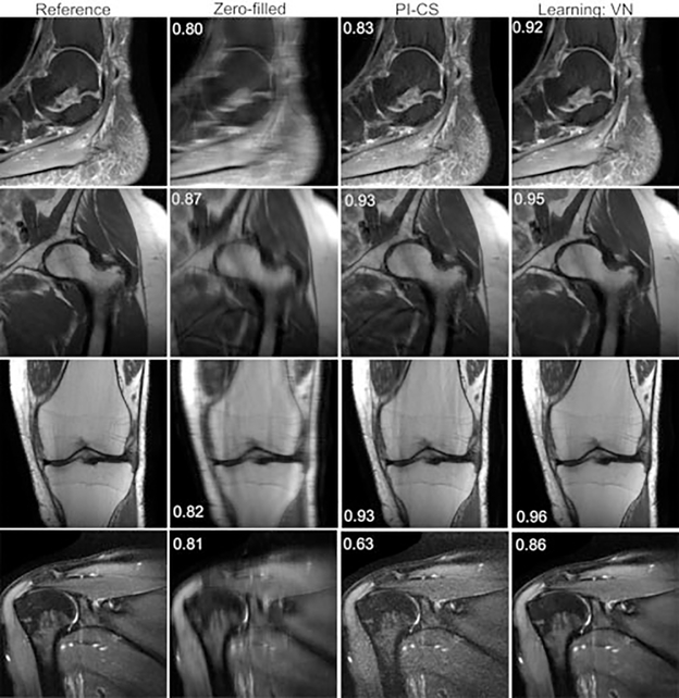
Fig. 8.
Ankle (sagittal proton-density fat-suppressed [PD-FS]), hip (coronal PD), knee (coronal PD), and shoulder (T2-weighted FS) reconstructions with fourfold acceleration. The learned reconstructions appear sharper and have fewer residual artifacts than the parallel imaging compressed sensing (PI-CS) reconstructions. The displayed structural similarity index measure values were calculated for the presented slices.

