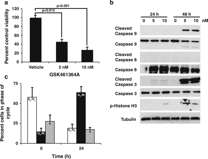
Fig. 1.
GSK461364A inhibits cell viability by causing cell cycle arrest and inducing apoptosis. a 231-BR cells were treated with 5 and 10 nM GSK641364A, respectively, for 48 h and viability assessed by MTT assay. Mean ± the standard error for data combined from three independent experiments shown as percent of vehicle control viability. A mixed model one-way analysis of variance (ANOVA) was performed on log10(y) data (P = 0.002) and results of the post-hoc Dunnett’s multiple comparison shown. b Western blot analysis of 231-BR cells treated with GSK641364A. Cells were treated for the times indicated with either 5 or 10 nM GSK641364A and cell lystates subjected to SDS-PAGE for standard western blotting. All antibodies used with the exception of Tubulin were from Cell Signaling Technologies. Tubulin (Calbiochem) shown as a protein loading control. c Cell cycle analysis was determined by FACS analysis after 231-BR cells were treated with 5 nM GSK461364A for 24 h. Data shown as percentage of cells in each phase of the cell cycle at the given time point. The mean ± standard deviation of data combined from two experiments is presented. White bars G0/G1 phase, Black bars G2/M phase, Grey bars S phase

