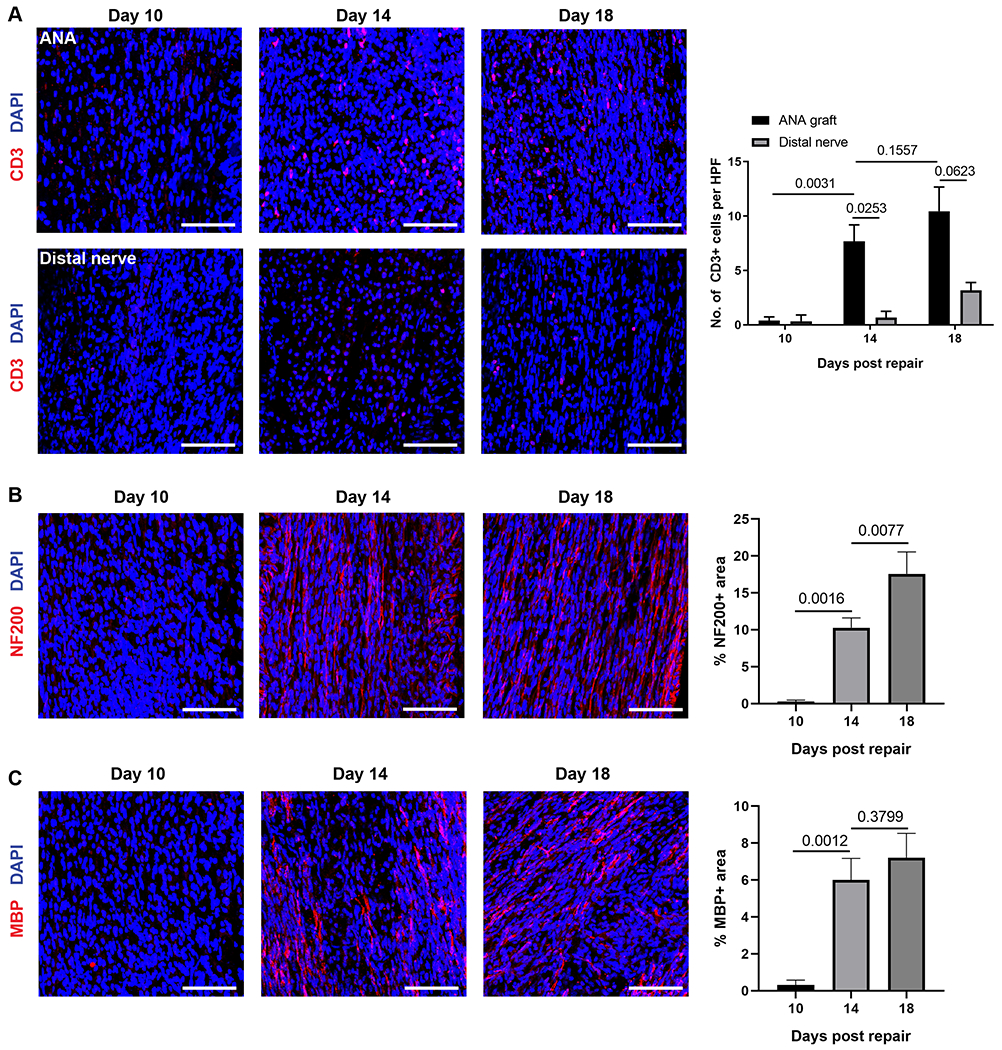
Fig 1.

T cells temporally accumulate within ANAs repairing sciatic nerve of WT mice. A) Representative immunofluorescence images of T cells (CD3, red) within ANA and distal nerve at 10, 14, and 18 days after repair with corresponding quantification. B) Representative immunofluorescence images of axons (NF200, red) within ANA and at 10, 14, and 18 days after repair with corresponding quantification. C) Representative immunofluorescence images of myelinated axons (MBP, red) within ANA and at 10, 14, and 18 days after repair with corresponding quantification. Mean ± SD, n=3/group. Scale bar represent 50 μm.
