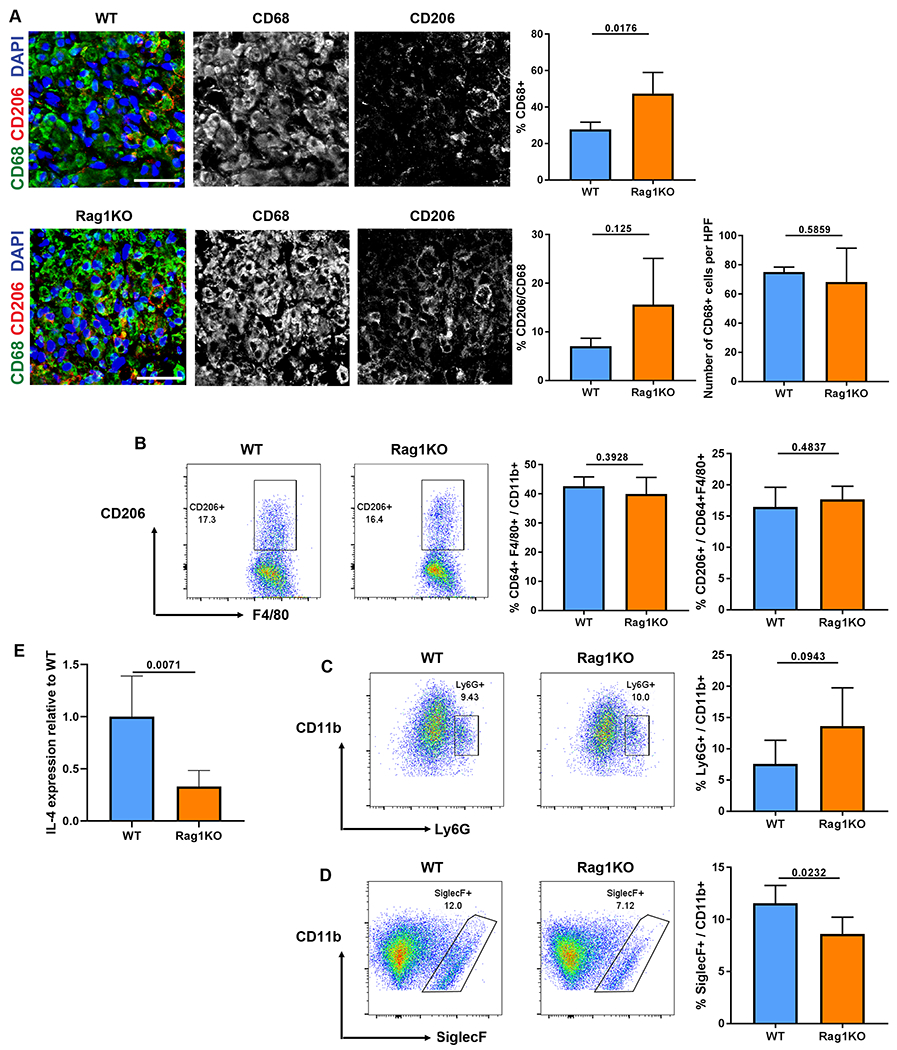
Fig 5.

Cytokine expression and myeloid cell repopulation within ANAs is altered in Rag1KO mice at 2 weeks. A) Representative immunofluorescence and quantification of macrophages (CD68, green) and CD206 macrophages (CD206, red) from the mid-graft of ANAs. B) Representative flow cytometry and quantification of macrophages (F4/80 CD64) among myeloid cells (CD11b), and proportion of CD206 macrophages among all macrophages. Cells were gated on CD45+CD11b+F4/80+CD64+.. Representative flow cytometry and quantification of neutrophils (Ly6G, C) and eosinophils (Siglec-F, D) among myeloid cells. Cells were gated on CD45+CD11b+. E) Gene expression of IL-4 from all cells contained within ANAs. Mean ± SD, n=5/group; p values shown. Scale bar represent 50 μm.
