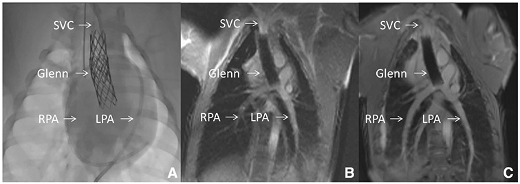
Fig. 1.

X-Ray Fluoroscopy versus Interventional Cardiac MRI (iCMR). This figure demonstrates a novel percutaneous intervention: transcatheter cavopulmonary anastomosis [58]. a Shows traditional X-Ray fluoroscopy of the endograft based cavopulmonary anastomosis (Glenn) in situ. Superior Vena Cava (SVC), Right and Left Pulmonary Artery (RPA, LPA) expected locations are shown. b Shows real-time CMR fluoroscopy with superior anatomic visualization. SVC, RPA, LPA and entire thoracic context are clearly and continuously displayed. c Shows higher quality diagnostic CMR (steady-state free precession “white” blood imaging) of the Glenn. During iCMR cases, intermittent higher quality non-real-time CMR can be obtained at any time
