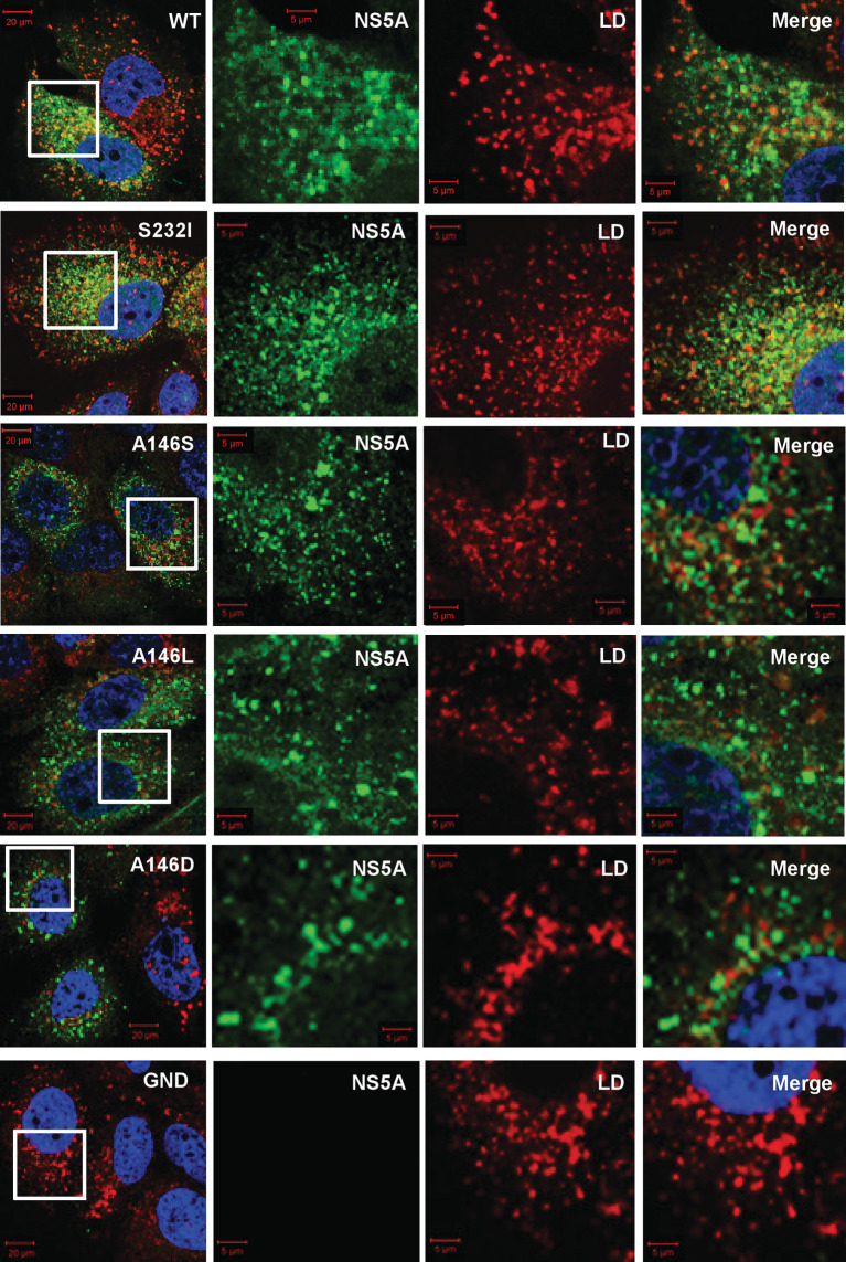
Fig. 3.
Sub-cellular distribution of NS5A and lipid droplets (LDs) in Con1 SGR electroporated cells. Huh7.5 cells were electroporated with in vitro transcripts of the indicated SGRs, and the cells were seeded onto coverslips and incubated for 48 h prior to fixation. Cells were subsequently permeabilized and immunostained for NS5A (sheep anti-NS5A), with LD detection using BODIPY(558/568)-C12 dye, prior to imaging by confocal microscopy. White boxes indicate the area expanded in the right-hand panels.

