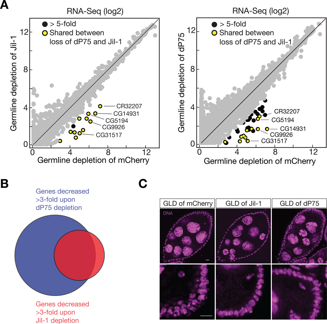
Fig. 6.
dP75 and Jil-1 function together to ensure oogenesis. A: Expression of genes measured by RNA-Seq. Gene expression is calculated as FPKM, and both the x-axis and y-axis are in log2 scale. B: dP75 and Jil-1 protect common targets for transcription. C: DAPI staining shows similar defects of germ cells by either depleting of dP75 or Jil-1 in germ cells. Upper panel: arrow heads point the nurse cells failed to pass the five-blob configuration. Percentage of each panel: 100% in GLD of mCherry (normal morphology); 57% in GLD of Jil-1 (abnormal morphology); 71% in GLD of dP75 (abnormal morphology). Stage 7 to stage 9 egg chambers were observed for quantification. Lower panel: oocyte DNA failed to compact into karyosome (pointed by arrow head). Percentage of each panel: 98% in GLD of mCherry (normal morphology), 34% in GLD of Jil-1 (abnormal morphology), 27% in GLD of dP75 (abnormal morphology). Stage 7 to stage 8 egg chambers were observed for quantification.

