
Fig. 3 |.
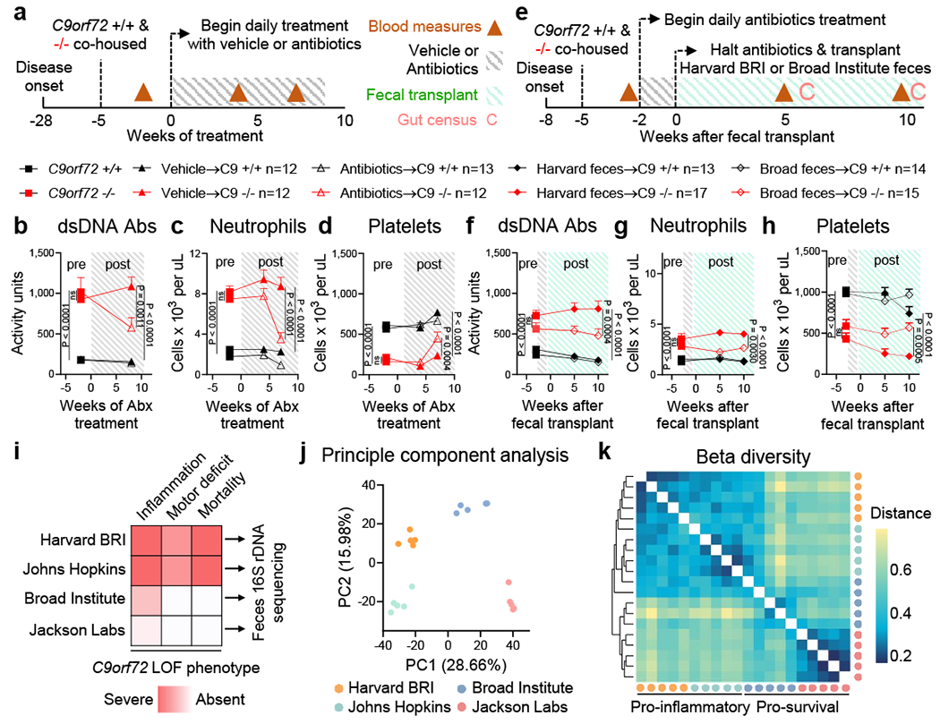
Gut bacteria propagates inflammation and autoimmunity in C9orf72 LOF mice
a, Age matched (36-week-old) female C9orf72Harvard +/+ and −/− neo deleted mice were cohoused by treatment group, then administered vehicle (+/+ n=12; −/− n=12) or antibiotics (+/+ n=13; −/− n=12) daily and assessed for b, plasma anti-dsDNA antibody activity, c, blood neutrophil count and d, blood platelet count measured at 0ºC. b-d and f-h, One way ANOVA with Sidak multiple comparisons. Each dot represents one animal. e, Age matched (13-week-old) female C9orf72Harvard +/+ and −/− neo deleted mice were cohoused by treatment group, administered antibiotics for two weeks, then gavaged Harvard BRI feces (+/+ n=13; −/− n=17) or Broad feces (+/+ n=14; −/− n=15) and assessed for f, plasma anti-dsDNA antibody activity, g, blood neutrophil count and h, blood platelet count measured at 0ºC. i, Fecal pellets (n=5 each) from two pro-inflammatory environments (Harvard BRI/Johns Hopkins) and two pro-survival environments (Broad Institute/Jackson Labs) were subjected to 16S rDNA sequencing and assessed by j, principle component analysis and k, Bray-Curtis dissimilarity matrix of beta diversity.