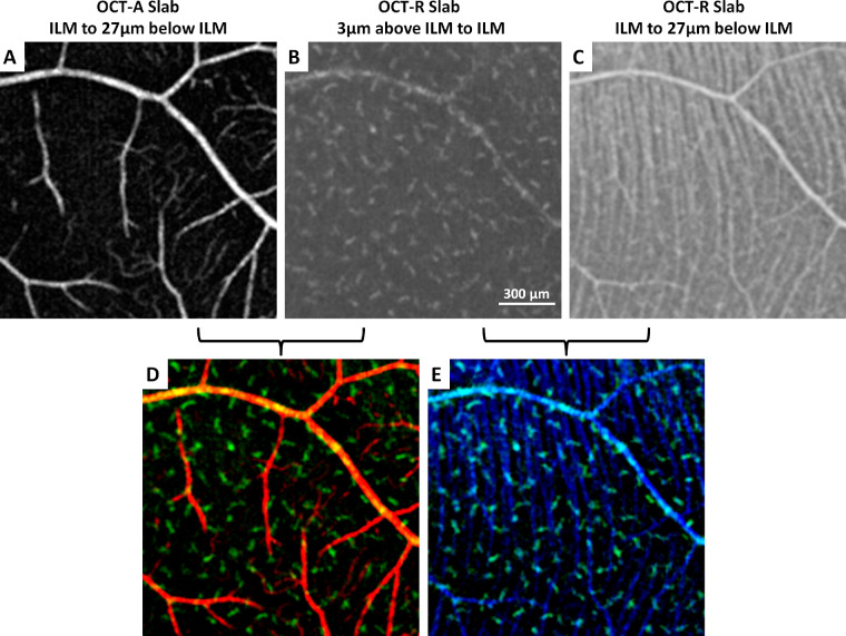
Figure 2.
Simultaneous imaging of (A) superficial retinal vascular network, (B) macrophage-like cells, and (C) RNFL at the temporal retina in a healthy control. (D, E) Overlay of superficial retinal vascular network (red), macrophage-like cells (green), and retinal nerve fiber bundles (blue) shows the spatial relationships among structures. Fovea is located to the left of all images.

