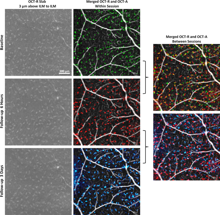
Figure 5.
In vivo imaging of macrophage-like cells over time in a healthy control (same subject as shown in Fig. 4). Left column: 3-µm OCT-R slabs located above the ILM surface at the temporal retina. Middle column: OCT-R (baseline in green, 6 hours follow-up in red, and 3 days follow-up in blue) and OCT-A overlaid. Right column: Merged OCT-R and OCT-A images from two imaging sessions. Cell structures are in different locations at 6 hours and 3 days later, suggesting cell translocation over time. Fovea is located to the left of all images.

