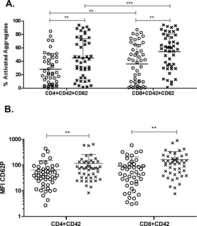
Fig 3. Platelet activation within platelet-T cell aggregates in lung cancer patients compared to healthy volunteers.
Whole blood from healthy volunteers or lung cancer patients was labeled with markers for CD4+ T cells (anti-CD4) or CD8+ T cells (anti-CD8) and were co-labeled for platelets (anti-CD42b) and activated platelets (anti-CD62P, P-selectin). Data was collected by flow cytometry. Populations were gated based on CD4+ and CD8+. PTCAs were identified as in Fig 2. Populations were further gated based on PTCAs and the percent of activated platelets (A) and MFI of activated platelets (B) within PTCAs were calculated. Mann Whitney nonparametric test. Error bars represent mean ± SD. N = 44–52. ** p ≤ 0.01, *** p ≤ 0.001.

