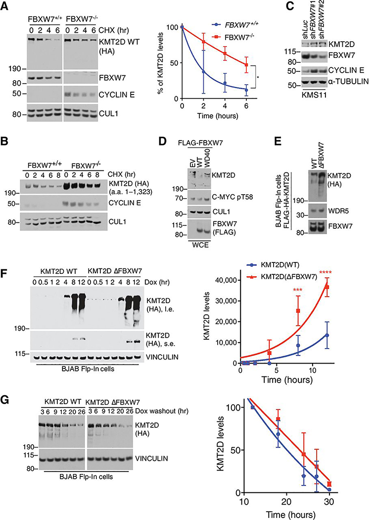
Figure 4. FBXW7 promotes degradation of KMT2D.
A, Immunoblot for the indicated proteins from HEK293T cells (FBXW7+/+ or FBXW7−/− CRISPR clones) overexpressing HA-KMT2D WT full-length and treated with cycloheximide (CHX) for the indicated times. ImageJ quantification of band intensity is plotted in the graph (n=3; mean ± SD; one-tailed Student’s t-test (*p<0.05)). KMT2D half-life is 1.9h and 5.5h in the FBXW7+/+ and FBXW7−/− cells, respectively B, Same as in (a), except that indicated HA-KMT2D fragment was expressed. C, Immunoblot for the indicated proteins from KMS11 multiple myeloma cells infected with lentiviruses encoding the indicated shRNAs. D, Immunoblot for the indicated proteins in HEK293T cells overexpressing the indicated FLAG-FBXW7 constructs. E, Immunoblot analysis for the indicated proteins from Flp-In™ T-Rex™ BJAB DLCBCL cells stably expressing KMT2D(WT) or KMT2D(∆FBXW7) upon doxycycline treatment (1 μg/mL). F, Immunoblot for indicated proteins of Flp-In™ T-Rex™ BJAB cells. Cells were treated with doxycycline (1 ng/mL) for the indicated times. Quantification of HA-KMT2D signal was done using ImageJ software (n=3, mean ± SD, two-way ANOVA (***p<0.0002, ****p<0.0001)). G, Immunoblot for indicated proteins of Flp-In™ T-Rex™ BJAB cells. Cells were treated with doxycycline (1 ng/mL) for 4 h. Doxycycline was washed-out and cells harvested at the indicated times. Quantification of HA-KMT2D signal was done using ImageJ software (n=3, data are mean ± SD, two-way ANOVA).

