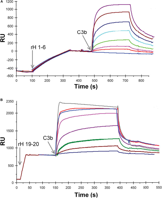
Figure 3.
SPR analysis of the binding of recombinant Factor H to C3b. (A) Purified rH 1-6 was attached to the NTA-Ni-coated sensor surface by injecting 20 μl of protein (1.2 μg/ml, in HBS-P) at a flow rate of 5 μl/min over flow cell 1. Eight different concentrations of C3b (0, 0.014, 0.028, 0.055, 0.11, 0.22, 0.44, and 0.88 μM C3b) in HBS-P were injected at 5 μl/min over flow cells 1 and 2. Lower concentrations of the ligand (C3b) were used than in (B) due to the higher affinity between rH 1-6 and C3b. (B) Same as in (A) except that purified rH 19-20 was attached to the NTA-Ni surface by injecting 20 μl of protein (3 μg/ml, in HBS-P) at a flow rate of 20 μl/min over flow cell 1. Eight different concentrations of C3b (0.14, 0.28, 0.57, 1.1, 12.0, 2.8, 4.3, 5.7 μM C3b) in HBS-P were injected at 5 μl/min over flow cells 1 and 2 (reference cell with Ni-NTA but no rH 19-20). RU (response units) shown represent the signal from flow cell 1 minus that from flow cell 2. The overlay plot was constructed using BIA Evaluation software. After each concentration of C3b was injected, the flow cells were regenerated with 300 mM imidazole.

