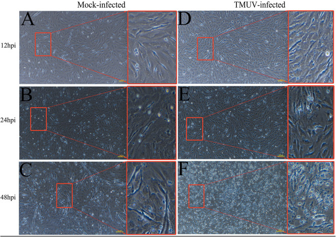
Figure 1.
Cytopathic effects of duck embryo fibroblast cells following DTMUV infection at 12, 24, and 48 h post-infection. Each sample had three biological replicates. The yellow scale bar represents 100 μm. (A) The status of mock-infected DEFs at 12 hpi. (B) The status of mock-infected DEFs at 24 hpi. (C) The status of mock-infected DEFs at 48 hpi. (D) The status of TMUV-infected DEFs at 12 hpi. (E) The status of TMUV-infected DEFs at 24 hpi. (F) The status of TMUV-infected DEFs at 48 hpi.

