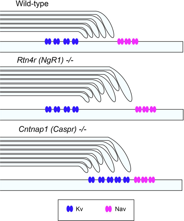
Figure 2.

Unaltered ion channel distribution in the axo-myelinic junction of the CNS of ngr1−/− mice. Schematic representation of the proposed ion channel distribution in the CNS myelinated fibers of wild-type, ngr1−/− and caspr−/− mice. In caspr−/−, juxtaparanodal Kv channels are displaced and distributed throughout the paranodal region, whereas in ngr1−/−, although lengthening of paranodal Caspr was found, ion channel distribution could not be identified (Lee et al., 2017).
