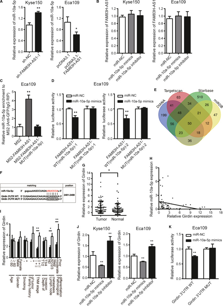
FIGURE 6.

FAM83H‐AS1 sponges miR‐10a‐5p and miR‐10a‐5p directly targets Girdin in oesophageal cancer cells. A, Relative expression of miR‐10a‐5p in FAM83H‐AS1 knockdown or overexpression cells. B, Relative expression of FAM83H‐AS1 in miR‐10a‐5p mimics or inhibitor transfected cells. C, The MS2‐RIP method identified the direct binding between FAM83H‐AS1 and miR‐10a‐5p. D, The effect of miR‐10a‐5p mimics on luciferase activity of wild‐type and mutant‐type FAM83H‐AS1 vectors observed by dual‐luciferase reporter assay. E, The numbers of miR‐10a‐5p targeting the potential same genes (including Girdin) drawn by Venn diagram. F, Schematic representation of the potential binding sites of miR‐10a‐5p on Girdin 3′ UTR. G, Relative expression of Girdin in 67 pairs of ESCC tissues and corresponding normal tissues confirmed by qRT‐PCR method. H, The correlation between Girdin and miR‐10a‐5p expression. I, Relative expression of Girdin in different subgroups. J, The regulation of miR‐10a‐5p on Girdin expression detected by qRT‐PCR method. K, The effect of miR‐10a‐5p mimics on luciferase activity of wild‐type and mutant‐type Girdin 3′ UTR vectors observed by dual‐luciferase reporter assay. Data are shown as mean ± SD; *P < .05 and **P < .01
