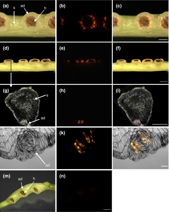
FIGURE 2.

Transformation of C. reflexa adhesive disk cells by A. rhizogenes containing the binary vector pRedRoot. (a‐f) Intact infection sites after transformation. Topview (a‐c) and sideview (d‐f) of transformed adhesive disks are shown. (g‐l) Semi‐thin vibratome sections of transformed adhesive disk tissue in the region of the stippled line (in d) show subepidermal localization of transformed cells in the adhesive disk (ad). (m, n) Mock transformation with A. rhizogenes lacking the binary pRedRoot plasmid. Darkfield or brightfield pictures (first column) are shown alongside the fluorescence images taken with a Cy3 filter (middle column). Overlays of both are shown in the right column. Adhesive disks (ad), haustoria (h), and stems (s) are indicated by arrows. Scale bars are 1,000 µm (c and i), 2,000 µm (f and n), and 100 µm (l)
