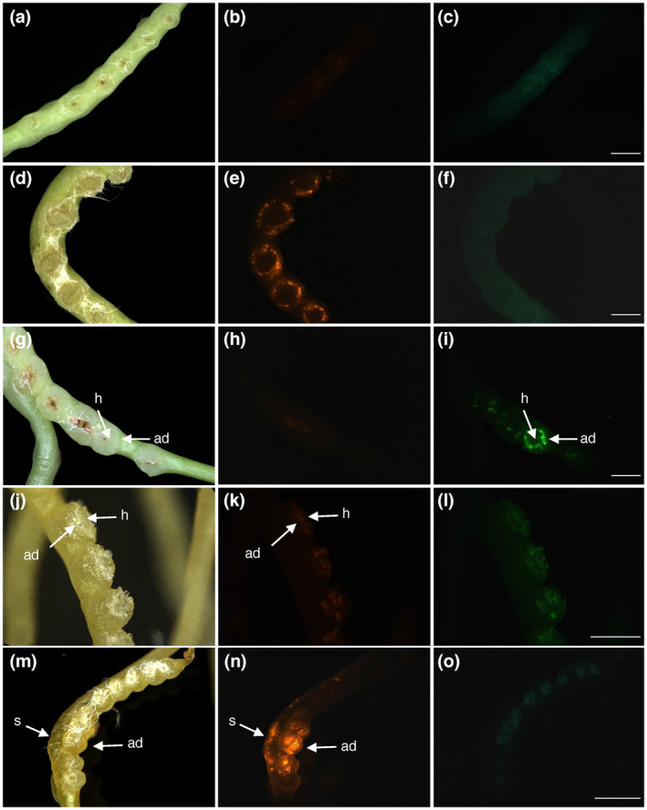
FIGURE 4.

Extension of the protocol to A. tumefaciens and C. campestris. (a–f) Negative (a–c) and positive (d–f) controls using the combination of C. reflexa and A. rhizogenes (see also Figure 2). (g–i) Transformation after combining C. reflexa with A. tumefaciens containing a binary GFP‐expressing vector. (j–o) Negative control (j–l) and pRedRoot transformation (m–o) using the combination of C. campestris and A. rhizogenes. Scale bars represent 2,000 µm (c, f, i and o) and 1,000 µm (l), respectively. White fibers of the bench paper from the experimental setup can be seen adhering strongly to the adhesive disks in some darkfield images (d, g, j and m)
