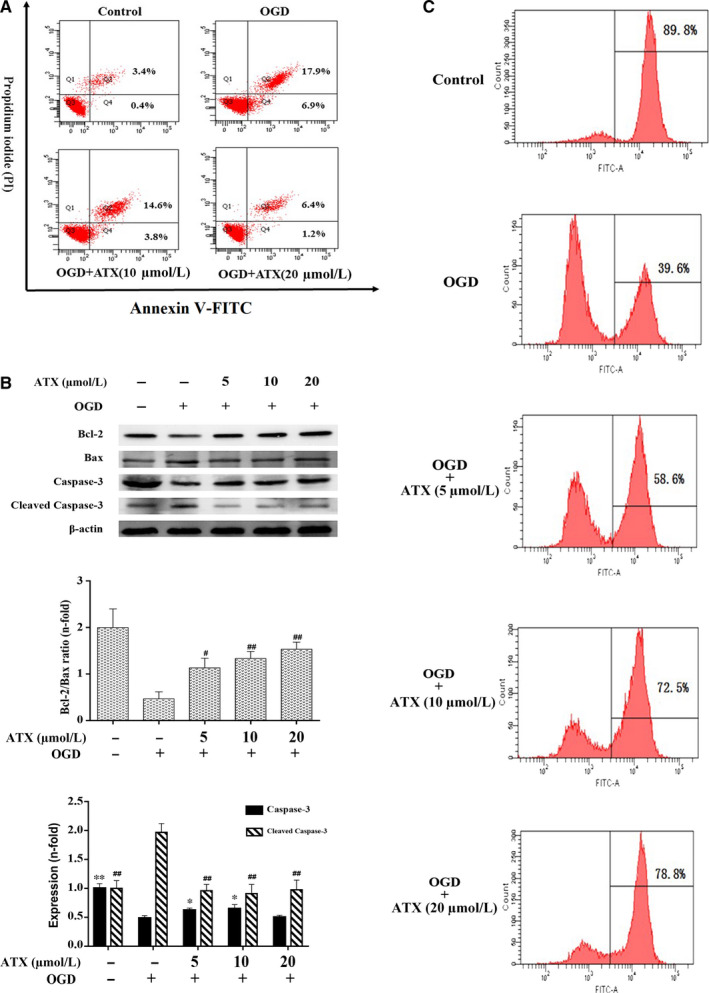
FIGURE 4.

Effect of ATX on the apoptosis of SH‐SY5Y cells. A, Apoptotic induction was determined by Annexin V/PI double‐staining assay after ATX treatment (10 and 20 μmol/L) for 24 h. B, Bcl‐2 Bax, caspase‐3 and cleaved caspase‐3 protein expressions were obtained by Western blot analysis. C, Cells treated with ATX (5, 10 and 20 μmol/L) for 24 h prior to exposure to OGD for 3 h were incubated with rhodamine 123 and evaluated by flow cytometry. The cell percentages in the right section of fluorocytogram indicated the number of mitochondrial membrane potential (Δψm)‐collapsed cells. Results were expressed as mean ± SD (n = 3). * #P < .05, ** ##P < .01 vs OGD group. Significance was determined by one‐way analysis of ANOVA followed by Dunnett's test
