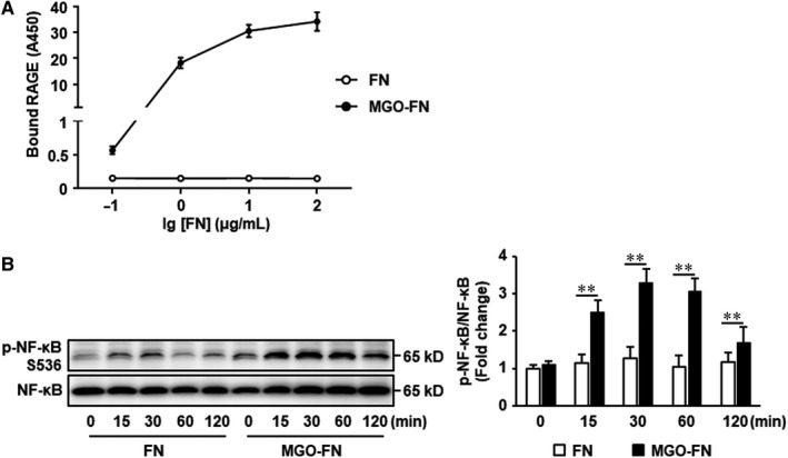
FIGURE 4.

Glycated FN directly binds to RAGE. A, 96‐well plates were coated with FN or MGO‐FN at 4°C overnight. After washing and blocking, recombinant RAGE (5 μg/mL) was added and incubated at 37°C for 1 hour. Bound RAGE was detected by sequential addition of biotinylated anti‐RAGE antibody, streptavidin‐HRP conjugate and HRP substrate and measurement of OD450. Data shown are mean ± SD for triplicate experiments. B, HUVECs were seeded onto FN or MGO‐FN for 0, 15, 30, 60 and 120 minutes. Phosphorylation (p) of NF‐κB and total NF‐κB were analysed by Western blotting in total cell lysates. Representative images of three independent experiments and densitometric analysis of phosphorylated NF‐κB normalized to total NF‐κB are shown. All data shown are mean ± SD for triplicate experiments and are expressed as fold changes. **P < .01
