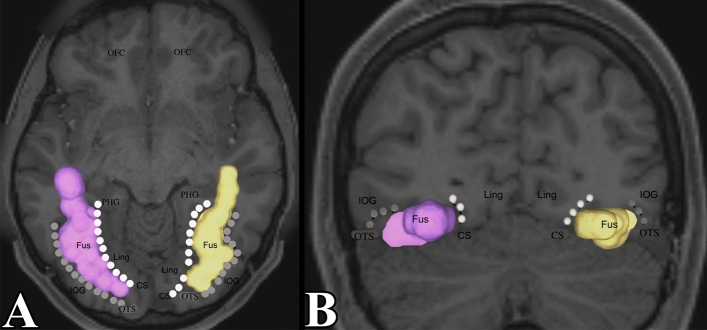
Figure 2.
The fusiform gyrus region of interest used in tractography. (A) Demonstrates an axial section of the fusiform gyrus. The collateral sulcus and occipito-temporal sulcus were identified to bound the fusiform gyrus from a medial–lateral direction. It was extended towards the temporal pole. We see that the fusiform gyrus does not reach the posterior-most aspect of the occipital lobe. (B) Demonstrates a coronal section of the fusiform gyrus. CS (collateral sulcus) = white dots, PHG = parahippocampal gyrus, Fus = fusiform gyrus, OTS (occipitotemporal sulcus) = grey dots, IOG = inferior occipital gyrus, ITG = inferior temporal gyrus, OP = occipital pole, OFC = orbito-frontal cortex.

