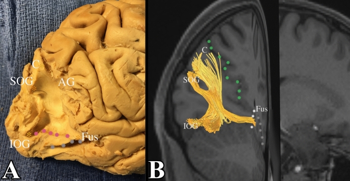
Figure 5.
Vertical occipital fasciculus. (A) Demonstrates a dissection deep into the white matter core of the occipital lobe. We first exposed connections between the superior occipital gyrus and the inferior occipital gyrus. Note how the fibers course obliquely from the superior occipital gyrus to the inferolateral occipital cortex (pink dots form the superior aspect the inferior occipital gyrus). The gross dissection represents the lateral portion of the VOF. We subsequently followed this back to expose a subcomponent of the VOF between the cuneus and fusiform gyrus. This is confirmed in tractography in (B). POS (parieto-occipital sulcus) = green dots, C = cuneus, CS (collateral sulcus) = white dots, Fus = fusiform gyrus, SOG = superior occipital gyrus, IOG = inferior occipital gyrus, OTS – grey dots, LOS (lateral occipital sulcus) = pink dots.

