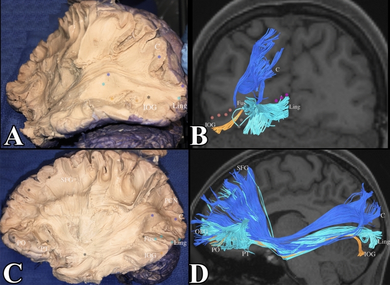
Figure 6.
(A) Demonstrates a dissection of the IFOF. Notice its subcomponents from the cuneus (demarcated in blue), lingual gyrus (light blue), and the inferior occipital gyrus (orange). This is confirmed in tractography in (B). The lateral occipital sulcus was included in pink dots to localize the fibers in the inferior occipital gyrus. The calcarine sulcus was included in purple dots to localize the lingual gyrus. We can see that subcomponents of the IFOF from the lingual gyrus and inferior occipital gyrus meet in the fusiform gyrus (black star) and course through the fusiform gyrus to reach the temporal lobe and their final destination in the frontal lobe. (C,D) Demonstrate the full IFOF. C = cuneus, L = lingual gyrus, IOG = inferior occipital gyrus, LOS (lateral occipital sulcus) = pink dots, CS (collateral sulcus) = purple dots.

