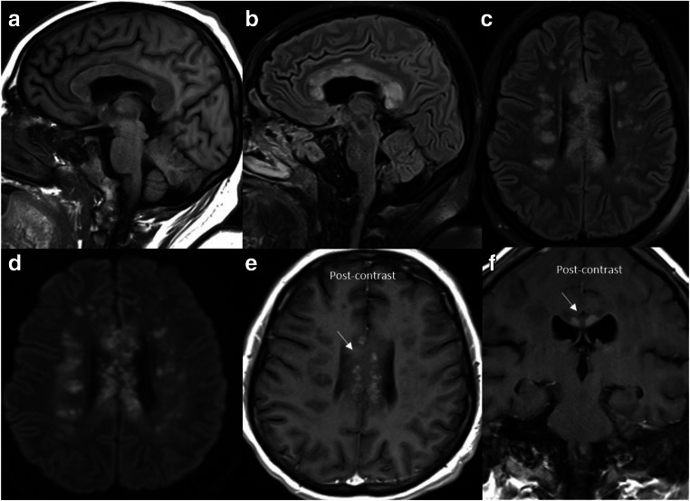
Fig. 3.
Acute tumefactive demyelination: Sagittal T1 (a), sagittal T2-FLAIR (b), axial T2-FLAIR (c), diffusion-weighted image (d), axial (e), and coronal (f) post-contrast images reveal extensive foci of demyelination in the supratentorial brain with marked involvement of the corpus callosum and pericallosal white matter. Many of these lesions show restricted diffusion and patchy enhancement (arrows)

