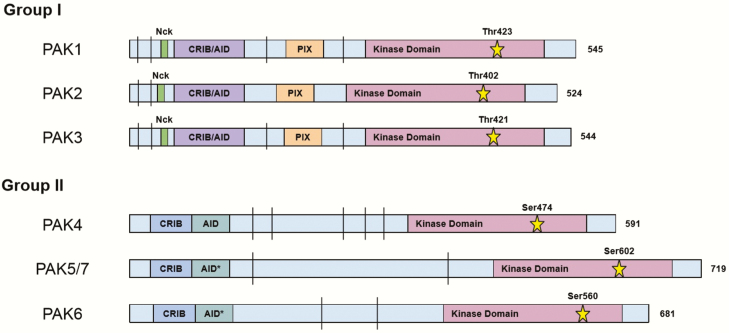
Figure 1.
PAK isoform structure. Linear representation of groups I and II PAK proteins, with the numbers to right indicating the total number of amino acid residues. Overlapping CRIB (CDC42/RAC interactive binding) domain, also known as the PBD, and AID (Auto-Inhibitory Domain) for group I PAKs is shown in purple. Separate CRIB domain and AID for group II PAKs are shown in blue and aquamarine, respectively. Nck and PIX binding regions are shown in green and orange, respectively. The kinase domain is shown in plum and the catalytic residue is represented by the star, with the corresponding residue number above. The vertical bar (|) indicates proline-rich segments that are potential binding regions for proteins with a SH3 domain. The asterisk indicates that the AID for these proteins, PAK5 and 6, may not actually serve as an inhibitory domain and needs further characterization. Drawing is not to scale.

