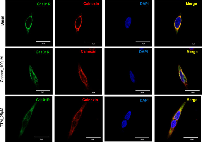
Figure 7.
Localization of G1101R ATP7B mutant in HEK293 cells under low and high Cu conditions. HEK293 cells were transfected (12 h) with WT or G1101R mutant ATP7B GFP-ATP7B (green) and treated for 3 h with basal medium (top), medium containing 100 µM of CuCl2 (middle), or medium containing 25 µM of tetra-thiomolybdate (TTM) (lower panel).Endoplasmic reticulum (ER) marker, Calnexin, in red and nuclei (DAPI) in blue. Co-localization is indicated in yellow. Green signals are from GFP fluorescence.

