
Figure 5. (Single column). Effect of FSH glycoform preparations on β-catenin phosphorylation in porcine granulosa cells.
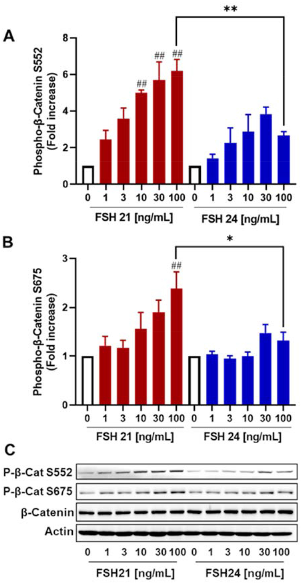
Cells were treated with increasing concentrations of hFSH21 or hFSH24 for 30 min. Cell lysates were prepared for Western blot analysis using phospho-Ser552 and phospho-Ser675-β-catenin (P-β-catenin) antibodies, and actin antibodies. A, fold changes in the phospho-β-catenin Ser552/Actin ratio; B, fold changes in the phospho-β-catenin Ser675/Actin ratio; C, representative Western blot analysis of phosphorylation of β-catenin after FSH glycoform treatment. Actin was used as an internal control. Data are expressed as means ± SEM; n = 3 separate experiments. #P < 0.05 and ##P < 0.01 compared with respective control; *P < 0.05 and **P < 0.01 compared with the other glycoform at the same concentration.