
Fig. 7.
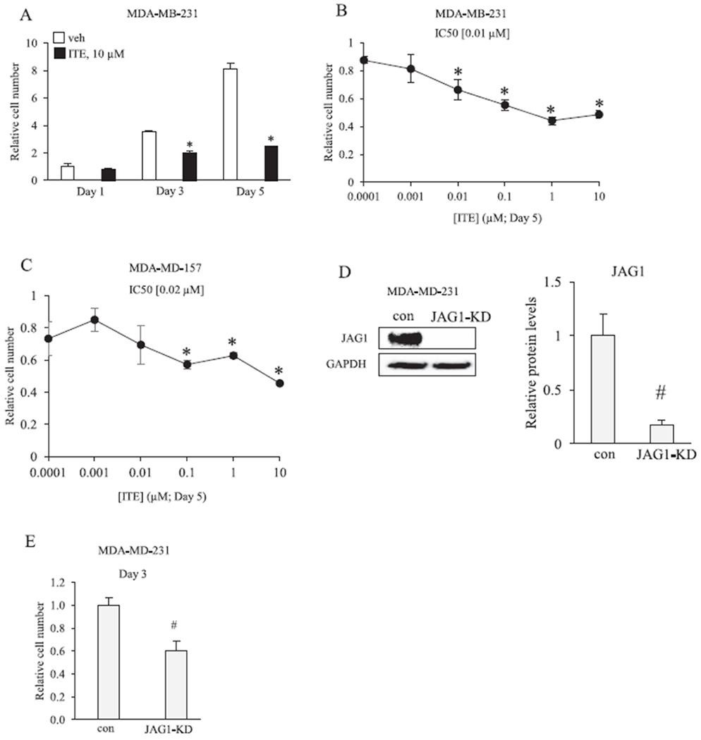
ITE reduces the growth of breast cancer cells. A, MDA-MB-231 cells were replenished with vehicle or 10 μM ITE every 12 h and the number of viable cells was determined by cell counting after 1, 3 and 5 days. B &C, Cells were replenished with increasing concentrations of ITE every 12 h and the number of viable cells was determined by cell counting on day 5. D, MDA-MB-231 cells were transfected with short interfering RNAs that were either non-targeting (con) or JAG1 targeting (JAG1-knockdown (JAG1-KD)). Total cellular extracts were collected and analyzed by western blot with JAG1 and GAPDH antibodies. E, The number of live MDA-MB-231 cells (con versus JAG1-KD) was determined on day 3 post transfection. Statistically significant changes induced by ITE, compared with vehicle controls treated for the same period of time, based on analysis of one-way ANOVA and Dunnett’s multiple comparison tests are indicated by *P < 0.05 (n = 3). Statistically significant changes induced by JAG1-KD compared with controls, based on Student’s t-test analysis, are indicated by #P < 0.05 (n = 3).