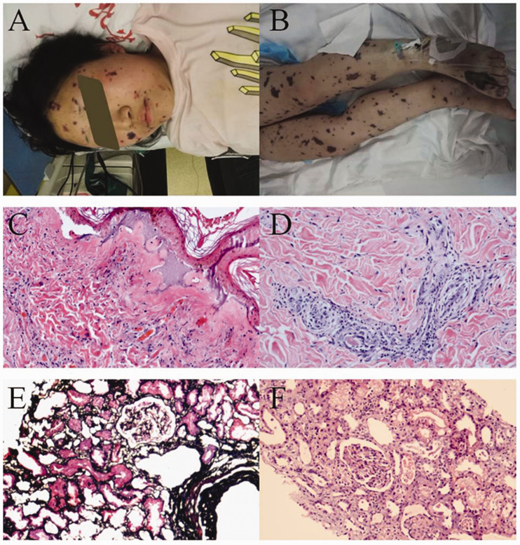
Figure 2.
Systemic ecchymosis and skin and renal pathologic findings of case 2. A and B. Extensive skin ecchymosis of the face and legs was observed in the patient with mTSS. C and D. Light microscopy examination of skin lesions showed massive accumulation of neutrophils and karyorrhexis within the dermis of the lesion center with wall necrosis in some of the small vessels and red blood cell overspill. Also visible was vascular endothelial swelling, narrowing of the vascular lumen and infiltration of the vascular wall by neutrophils and lymphocytes within the dermis surrounding the lesion. Subepidermal vesicles were observed as well. These changes were consistent with leukocytoclastic vasculitis. E and F. Light microscopy showed ischemic shrinkage of most glomerular capillary loops and, in the tubular epithelium, shedding of the brush border, lumen expansion, exposure of the basement membrane and epithelial regeneration. Exfoliated cell debris and proteins were observed within the lumen, slight edema of the interstitium and indiscernible changes within the arterioles. These alterations reflected acute tubular necrosis with ischemic injury.

