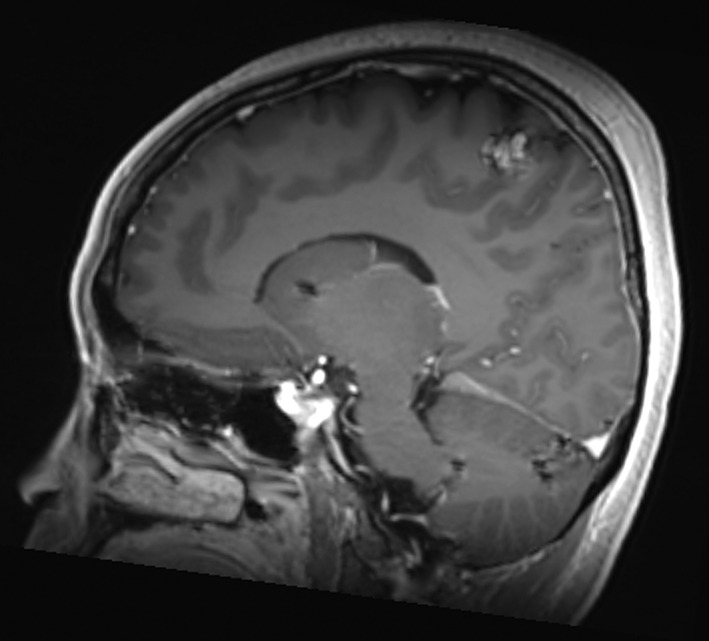
Figure 2.

Mrs. ND, July 2018 sagittal T1 FLAIR image with contrast of the 14‐mm right vertex lesion in the sensorimotor strip progressing while on third‐line alectinib. The remaining intraparenchymal lesions were unchanged, and no new lesions were appreciated.
