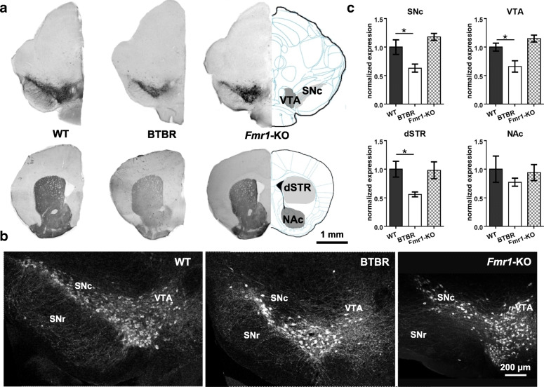
Fig. 1.
Immunohistochemical analyses of TH expression in WT, BTBR and Fmr1-KO mice. a Representative diagrams and images of anti-TH staining in substantia nigra pars compacta (SNc), ventral tegmental area (VTA), dorsal striatum (dSTR) and nucleus accumbens (NAc). b Examples of confocal images (20x) of dopaminergic neurons in WT, BTBR and Fmr1-KO mice. c Fluorescence intensity of anti-TH staining in the region of interest (ROI) was measured in an identical microscopic setting and normalized to the WT animals. The BTBR brain exhibited decreased TH-positive expression in SNc, VTA and dSTR, while the Fmr1- KO brain did not. TH: tyrosine hydroxylase; SNr: substantia nigra pars reticulata. n = 6–7 samples/group. *p < 0.05, compared to WT

