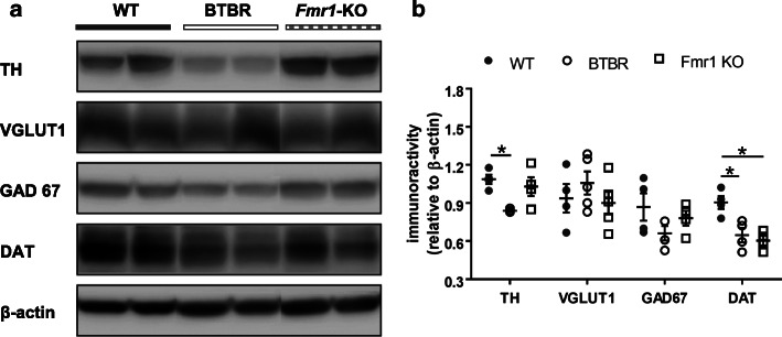
Fig. 3.
Western blotting of TH, VGLUT1, GAD67 and DAT in the striatum. a Examples of Western blots of striatal lysates from WT, BTBR and Fmr1-KO mice. Images of protein bands were aligned for comparison. b Blot intensity was normalized to an internal standard β-actin. Decreased TH and DAT levels were found in BTBR mice, while Fmr1-KO animals only showed reduced DAT expression, as compared to the WT group. Relative quantities of other proteins were comparable among groups. VGLUT1: vesicular glutamate transporter 1; GAD67: glutamate decarboxylase 67; DAT: dopamine transporter; TH: tyrosine hydroxylase. n = 4–5 mice/group. *p < 0.05, compared to WT

