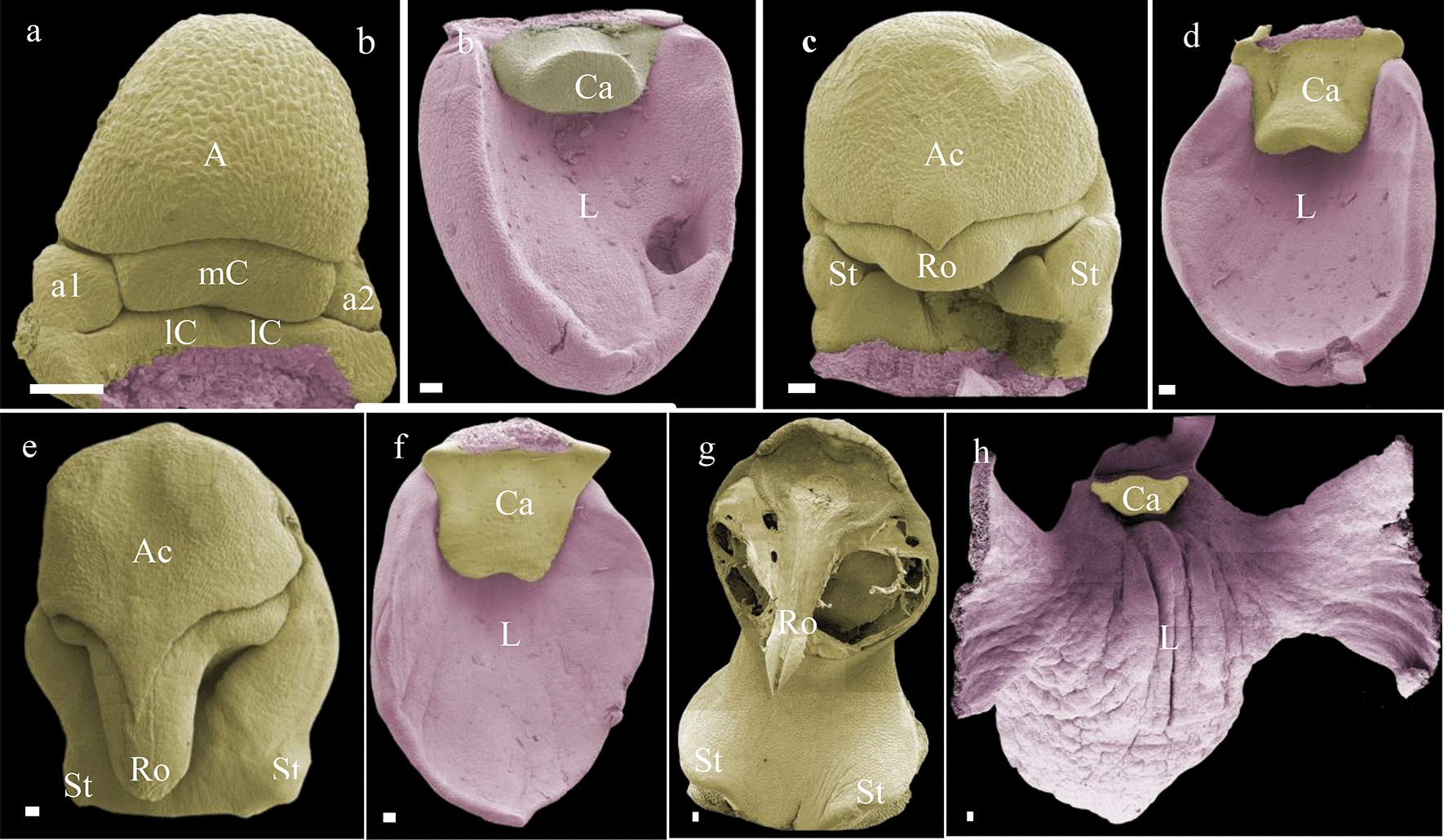
Fig. 5.

Floral ontogeny of Phalaenopsis pulcherrima. a Early stage gynostemium in which the staminodes are still visible as separate organs; b early formation of the callus on the labellum; c, d late stage floral bud in which the size of the anther cap, stelidia, rostellum, callus and labellum further increases; e almost fully developed apical part of the gynostemium, the rostellum pokes out from underneath the anther cap and the stelidia are almost fully merged with the gynostemium; f early stage labellum and almost fully developed callus; g late stage apical part of the gynostemium, from which the anther cap and pollinia were removed, showing the fully developed rostellum; h. fully developed labellum and callus. Images were made under various magnifications ranging from ×37 to ×130 magnification. A1 anther, a1–a2 staminodes, Ac anther cap, Ca callus, L labellum, lC lateral carpel, mC median carpel, St stelidia, Ro rostellum. Scale bar: 100 µm. Photographs by Dewi Pramanik
