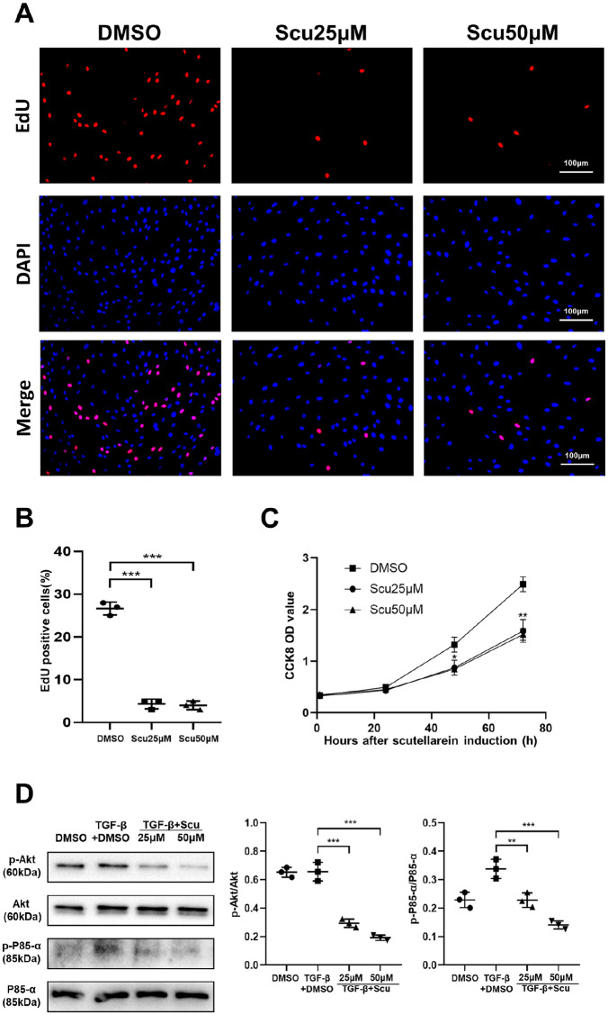
Figure 6.
Scutellarein inhibits fibroblast proliferation. (A) Typical image of EdU-stained HPFs following scutellarein stimulation. HPF was stimulated by scutellarein for 48 h. (B) Bar graph of the EdU staining results. (C) CCK8 proliferation curve. HPF was stimulated by scutellarein for 0h, 24 h, 48 h and 72 h. (D) Western blot analysis of p-Akt and p-P85-α. HPF was stimulated by TGF-β1 and scutellarein for 1 h. Left panel: representative western blot results. Right panel: bar graphs of the western blot results. The statistical figures show the data for three replications. The experimental results are expressed as the means ± standard deviations. The data were analyzed by one-way analysis of variance.
**p < 0.01; ***p < 0.001.
CCK8, Cell Counting Kit-8; EdU, 5-ethynyl-2′-deoxyuridine; HPF, human pulmonary fibroblast; Scu, scutellarein.

