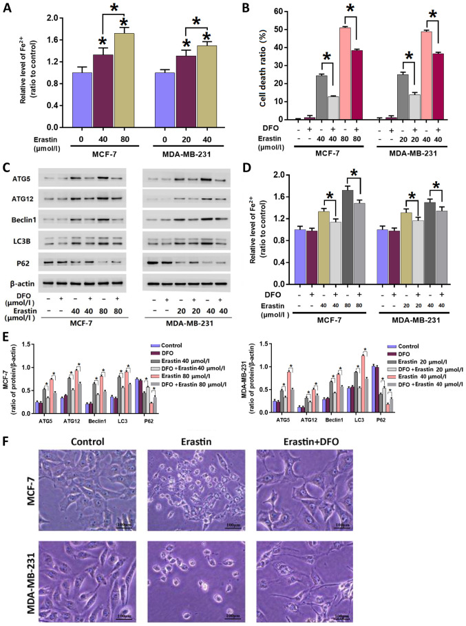
Figure 3.
Mitigation of erastin-induced increases in irons levels inhibits erastin-induced autophagy. (A) Erastin increased the levels of intracellular Fe2+ in MCF-7 and MDA-MB-231 cells in a dose-dependent manner. (B) LDH release assay demonstrated that the breast cancer cell death induced by erastin was inhibited in the presence of DFO. (C) Western blotting and (E) quantification demonstrated that the upregulation of autophagy-related protein expression levels induced by erastin was suppressed by DFO. (D) Pretreatment with 500 µmol/l DFO for 1 h mitigated the erastin-induced increases of intracellular Fe2+. (F) Representative light microscope images of MCF-7 and MDA-MB-231 breast cancer cells. The majority of cells treated with erastin alone became round in shape and smaller in size, which was prevented by pretreatment with DFO. Scale bar, 100 µm. *P<0.01 vs. untreated control group or as indicated. DFO, deferoxamine; LDH, lactate dehydrogenase; ATG, autophagy related; LC3B, microtubule-associated proteins 1A/1B light chain 3B.

