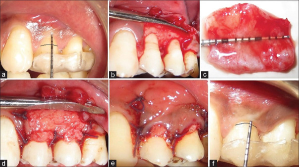
Figure 2.
Subepithelial connective tissue graft group. (a) Preoperative view Miller's Class I recession on teeth #12 and #13. (b) Flap is reflected. (c) Harvested connective tissue. (d) The connective tissue was trimmed to the shape of the recipient site and sutured in place. (e) The overlying flap was placed coronally to cover the graft and sutured. (f) 6-month postoperative view of the treated site

