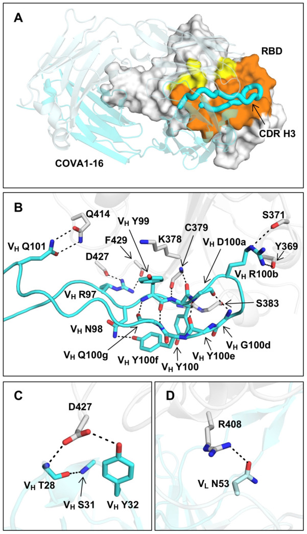
Figure 3. Interaction between SARS-CoV-2 RBD and COVA1–16.

(A) The epitope of COVA1–16 is highlighted in yellow and orange. Epitope residues that are in contact with CDR H3 are in orange, and yellow otherwise. COVA1–16 (cyan) is in cartoon representation with CDR H3 depicted in a thick tube. The RBD (white) is in a surface representation. The BSA on COVA1–16 and RBD are 844 Å2 and 779 Å2, respectively. (B) Interactions of SARS-CoV-2 RBD (white) with (B) CDR H3, (C) CDR H1, and (D) CDR L2 of COVA1–16 (cyan) are shown. Hydrogen bonds are represented by dashed lines. In (C), a 310 turn is observed in CDR H1 for residues VH T28 to VH S31.
