
FIGURE 8.
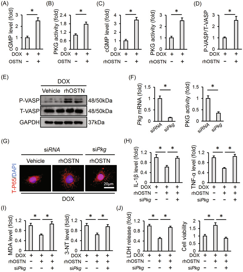
OSTN prevents DOX‐induced inflammation, oxidative stress, and apoptosis via activating PKG. A, Myocardial cGMP level with or without OSTN overexpression after DOX injection (n = 6). B, Relative PKG activity in the myocardium (n = 6). C, Relative cGMP level and PKG activity in DOX‐treated H9C2 cells with or without rhOSTN incubation for 24 h (n = 6). D and E, Representative western blot images and the corresponding statistical results in H9C2 cells with or without rhOSTN incubation (n = 6). F, H9C2 cells were incubated with siPkg (50 nmol/L) or siRNA (50 nmol/L) for 4 h and then maintained in normal medium for additional 24 h. The efficiency of siPkg in H9C2 cells was detected by western blot (n = 6). G, H9C2 cells were pre‐infected with siPkg or siRNA, and then received DOX insult (1µmol/L) with or without rhOSTN protection (5 µg/mL). Immunofluorescence staining of T‐P65 in H9C2 cells (n = 6). H, The releases of IL‐1β and TNF‐α from H9C2 cells to the medium (n = 6). I, The levels of MDA and 3‐NT in H9C2 cells (n = 6). J, LDH release from H9C2 cells and the quantitative data of cell viability (n = 6). Values represent the mean ± SD. * P < .05 versus the matched group