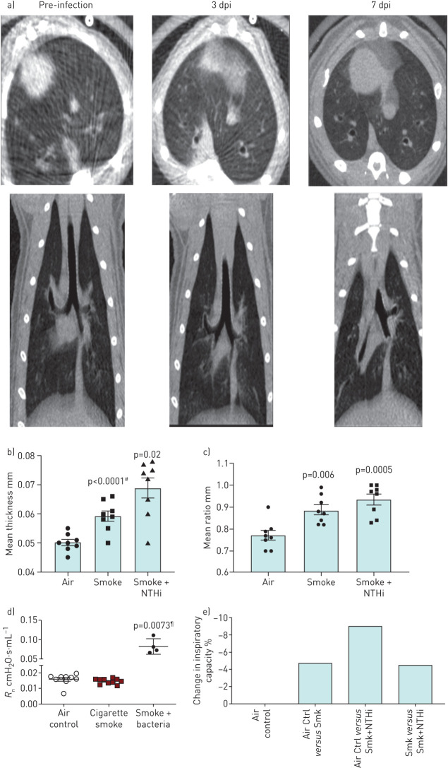
FIGURE 1.
Non-typeable Haemophilus influenzae (NTHi) infection induces significant morphological and functional decline in lung function in smoke-exposed ferret model of chronic obstructive pulmonary disease (COPD) indicative of an exacerbation event. Representative micro computed tomography (µCT) images of smoke-exposed ferret lungs infected with NTHi with quantification of airway walls. a) Axial view and posterior views of smoke-exposed ferret lungs prior to infection and post-infection at days 3 and 7. By day 3 post-infection (dpi) there is radiological evidence of consolidation (ground glass infiltrates), infection, and bronchial impaction, which is indicative of mucus build up in the bronchioles. b) Airway wall thickness and ratio of wall thickness to airway diameter of air controls, uninfected smoke-exposed ferrets, and infected smoke-exposed ferrets. #: comparison to air; ¶: comparison to smoke alone. Compared to air controls, smoke-exposed infected ferrets had significantly thicker airway walls and ratio of wall thickness to airway diameter indicating increased blockage and constriction. Mean±sem. n=8. c) Airway functional analysis of ferret airways. Central airway resistance is significantly increased in infected smoke-exposed animals compared to both smoke only and air controls as determined by one-way ANOVA. Mean±sem. n=4–10. Measurement of percent change in inspiratory capacity in pre- and post-infected smokers shows a synergistic effect between smoke exposure and bacterial infection.

