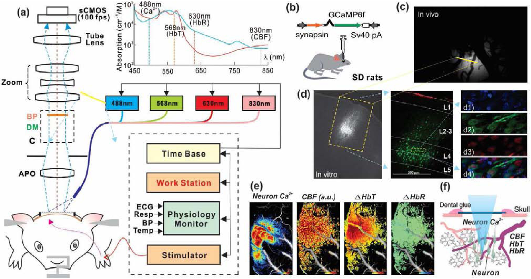
Figure 1.

A schematic diagram to illustrate the experimental approach. (a) Multimodality imaging platform that combines GCaMP6f Ca2+ fluorescence / spectral imaging and laser speckle contrast imaging for simultaneous detection of neuronal activity, cerebral metabolic and hemodynamic changes. Sensory stimulation (electrical forepaw stimuli) was synchronized with the imaging platform via a shared time base. (b) Viral injection to express GCaMP6f in neurons within somatosensory cortex. (c) In vivo image of neuronal Ca2+ signal from rat cortex. (d) Brain slice to show the GCaMP6f injection spot in the cortex at layer IV-V and confocal fluorescence images of brain slice with DAPI (d1), GCaMP6f (d2), NeuN (d3) and merged image (d4). (e) Simultaneous imaging of neuronal Ca2+, CBF, HbT and HbR responses to forepaw electrical stimulation (Image size: 3×5mm2). (f) Illustration of simultaneous imaging of synchronized Ca2+ from neuronal population and the local hemodynamics.
