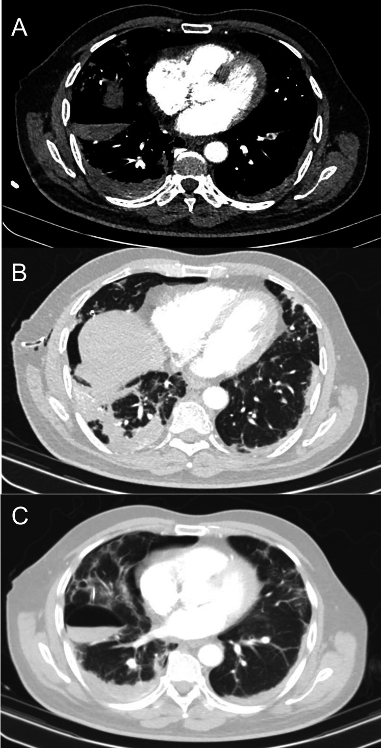
Figure 3.
CT pulmonary angiogram. (A) Segmental pulmonary emboli in the left lower lobe. (B) Generalised peripheral alveolar opacities in keeping with COVID-19 with dense bilateral lower lobe consolidation, more pronounced in the right lung. Chest drain is noted in the right axilla with residual right pneumothorax and subcutaneous surgical emphysema. (C) Iatrogenic secondary loculated pneumatocoele due to the chest drain traversing the lung parenchyma.

