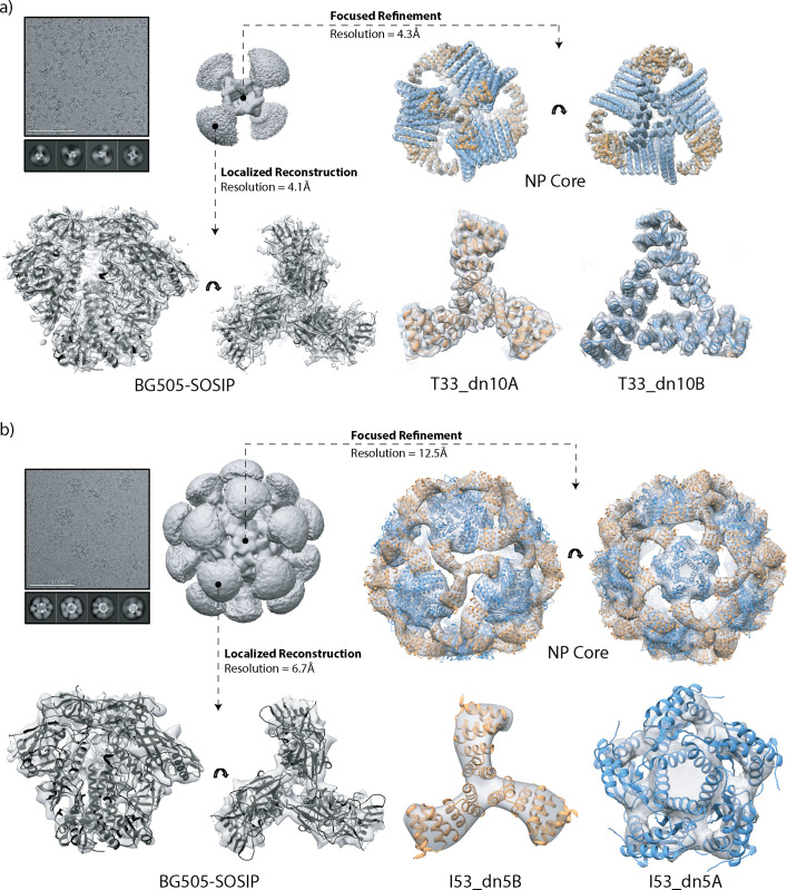
Fig 3. Cryo-EM analysis of tetrahedral and icosahedral nanoparticles.
(a) Tetrahedral BG505-SOSIP-T33_dn10 nanoparticle; (b) Icosahedral BG505-SOSIP-I53_dn5 nanoparticle. Sample micrograph, 2D class averages and initial 3D reconstructions of the full nanoparticles are displayed in the top left part of the corresponding panels. Focused refinement was applied to generate a 3D reconstruction of the nanoparticle core (top and bottom right, maps are in light gray). The refined model of T33_dn10 and the Rosetta_design model of I53_dn5 are docked into the corresponding maps (antigen-bearing component, orange; assembly component, blue). Localized reconstruction approach was used for analysis of the presented antigen (bottom left). Refined BG505-SOSIP models are shown in black.

