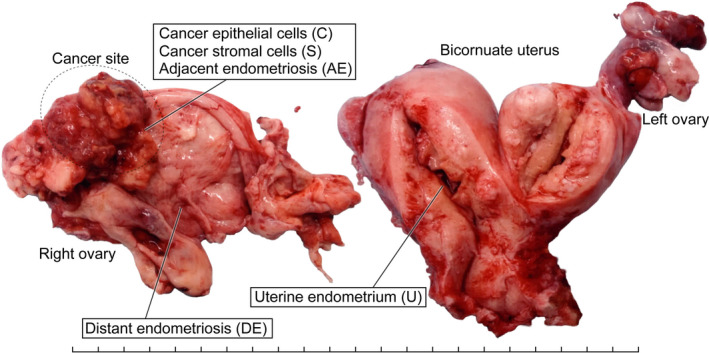
FIGURE 1.

A macroscopic picture of a surgical specimen with sampling sites. A, An image of a surgical specimen from a 56‐year‐old woman with ovarian clear cell carcinoma, representing multiregional sampling sites of the following tissue components: cancer epithelial cells (C), cancer stromal cells (S), epithelial cells of adjacent endometriosis (AE) and distant endometriosis (DE), and epithelial cells of uterine endometrium (U). Each tick mark represents 1 cm
