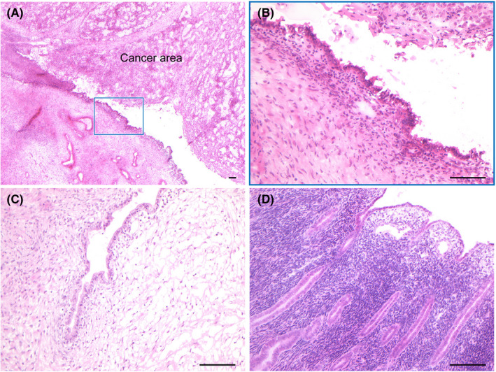
FIGURE 2.

Histological images of multiregional samples. Frozen sections of 8‐µm thickness from each sample were stained with H&E to histologically confirm (A) the cancer site accompanied by adjacent endometriosis (boxed lesion), (B) adjacent endometriosis, (C) distant endometriosis and (D) uterine endometrium. The scale bars represent 100 µm
