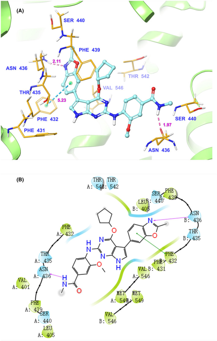
FIGURE 5.

Computational molecular docking binding of CC‐671 to the human ABCG2 model. The binding mode of CC‐671 to the human ABCG2 model predicted by induce‐fit docking. A, Docked conformation of CC‐671 (ball and rod model) is shown within the ABCG2 drug‐binding cavity, with the atoms colored as follows: carbon, cyan; hydrogen, white; oxygen, red; nitrogen, blue. The ABCG2 structure is in the ribbon diagram in light green. Important amino acid residues are described (rods model) with the same color scheme as above for all atoms but carbon atoms in orange. Dotted blue lines represent hydrogen‐bonding interactions, whereas dotted azure lines represent π‐π stacking interactions. The values of the correlation distances are indicated in Å. B, The 2‐D schematic diagram of ligand‐receptor interaction between CC‐671 and the human ABCG2 model. Amino acids within 4 Å are indicated as colored bubbles, polar residues are depicted as light blue, and hydrophobic residues are depicted as green. Pink arrows denote H‐bonds and dark green lines denote π‐π stacking aromatic interactions
