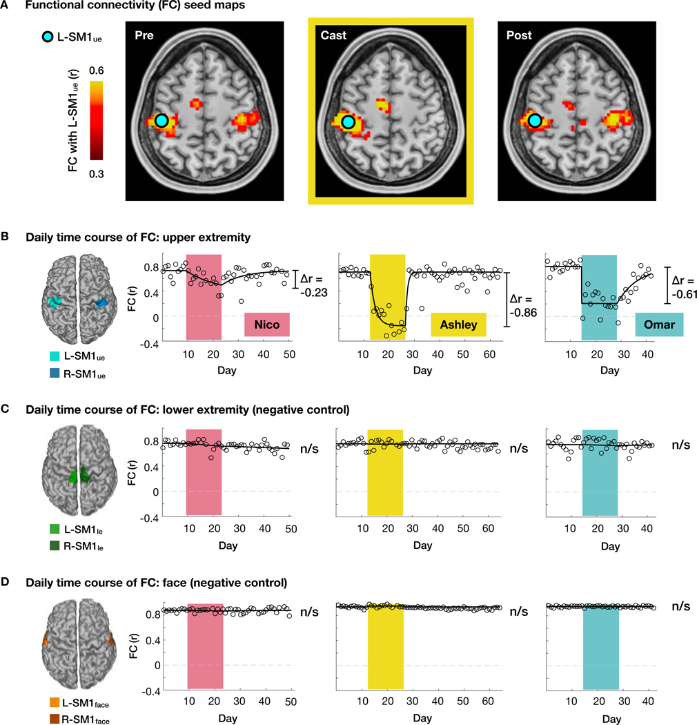
Figure 2. Disused somatomotor cortex became functionally uncoupled from opposite hemisphere.
(A) Seed maps showing functional connectivity (FC) of each voxel with the left primary somatomotor cortex (L-SM1ue) during scans acquired before, during and after casting (Pre, Cast, Post) in an example participant (Ashley). The L-SM1ue region of interest (ROI) was defined using task functional MRI. (B) Daily time course of FC between L-SM1ue and R-SM1ue for each participant. Δr values are based on a time-varying exponential decay model (black lines, dr⁄dt = α(r∞ − r); Nico: P = 0.002, Ashley: P < 0.001, Omar: P<0.001). (C and D) Daily time course of FC in lower extremity (C) and face (D) regions of the left and right somatomotor cortex.

