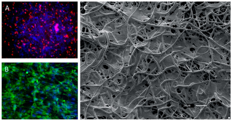
Figure 4.
Morphology of culture in the scaffold. (A) Fluorescence microscopy of adipose-derived stem cells (ASCs) staining with PKH26 in the first hours of culture in the scaffold; (B) fluorescence microscopy of culture of ASC in scaffold staining with phalloidin–FITC; (C) Scanning electron microscopy of ASCs on multi-polydioxanone (PDO) scaffold after 14 days of culture.

