Fig. 1.
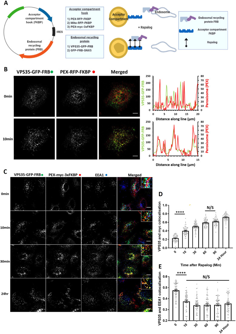
Knocksideways can rapidly mislocalise retromer from endosomes. (A) Schematic showing the design of the endosomal knocksideways system. (B) HeLa cells transfected with retromer knocksideways (PEX–RFP–FKBP and VPS35–GFP–FRB). Still frames are shown from a movie (Movie 1A) at either 0 min or 10 min after the addition of rapalog. Line scans were generated using ImageJ by drawing a line through peroxisome structures, and represent the colocalisation between VPS35–GFP–FRB and PEX–RFP–FKBP at each time point. The merged panel displays both channels. (C) Retromer knocksideways HeLa cells were fixed at multiple time points after the addition of rapalog. Anti-Myc and anti-EEA1 antibodies were used to label PEX–Myc–3×FKBP and early endosomes, respectively, and the merged panel displays triple colocalisation between three channels. Magnified images are displayed in the insets at the top right of the merged image. (D) Pearson’s colocalisation between VPS35–GFP–FRB and PEX–Myc–3×FKBP (peroxisomal targeting sequence) at multiple time points after the addition of rapalog. nexp=3, ncell=60 with all data points being displayed. (E) Pearson’s colocalisation between VPS35–GFP–FRB and EEA1 (early endosome marker) at multiple time points after the addition of rapalog. nexp=3, n=60 with all data points being displayed. ****P<0.0001; N/S, not significant (P>0.05) (ordinary one-way ANOVA with multiple comparisons). Error bars show the s.e.m. Scale bars: 10 µm.

