
Figure 4.
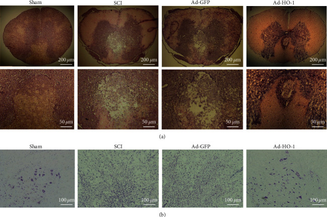
Observation of structural damages and neuronal necrosis. (a, b) The structural damages and neuronal necrosis were detected with the HE staining (a) and Nissl staining (b). The images were obtained at low microscope objective magnification of 5x, 20x, and 10x.