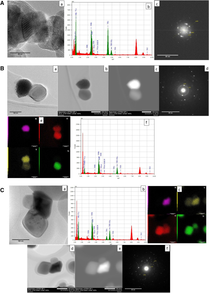
Figure 6.
A Micrographs of the 1:10 nanocomposite (a) TEM micrograph (b) EDS chemical analysis (c) FFT diffraction pattern. B Micrographs of the 1:2.5 nanocomposite (a) TEM micrograph of isolated particles (b, c) bright and dark field analysis (d) FFT diffraction pattern (e) elemental mapping and (f) EDS analysis respectively. C Micrograph of the nanocomposite 1:0.6 (a) isolated particles (b) EDS analysis of the nanocomposite (c) elemental mapping of the nanocomposite, (d) bright-field image (e) dark-field image (f) FFT diffraction pattern.

